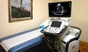
 Echocardiogram (ECHO), or Echocardiography, is a diagnostic test that uses sound waves, called ultrasound, to look at the shape, size, and motion of the heart, heart valves, the walls of the heart, and the blood vessels entering and leaving the heart. It is a non-invasive and painless test that can be performed in the office as the Sonographer puts a special jelly on a probe and gently moves it around over your chest area.
During the test, ultra-high-frequency sound waves simply pick up images of your heart and valves without the use of X-rays. The movements of your heart can then be viewed on a video screen and a videotape or a photograph can be made. The procedure typically takes about an hour and you can sometimes watch the screen as well during the test.
There are other types of Echocardiograms, and some require inserted tubes to gain further details and clearer images while others require for you to exert some energy to get your blood really flowing. These include: Transesophageal Echocardiogram, Doppler Echocardiogram, and Stress Echocardiogram.
The purpose behind having an Echocardiogram is to aid in the assessment of health within the structure and functioning of your heart. It is just one part of assessing and monitoring potential and established heart problems. There are no special preparations required for a standard Echocardiogram test, so you can eat and take medications as you would normally; however, your doctor will request that you not to eat for a few hours if your test is a Transesophageal or Stress Echocardiogram.
The test can evaluate the general size of your heart, the size of your four heart chambers, and the appearance and function of the four heart valves. It can also assess and view the two septa – the atrial heart septum separates the right and left atrium and the ventricular heart septum separates the right and left ventricles. An Echocardiogram can also assess the condition of the the sac that lines the heart called the pericardium, as well as the state of the aorta. It can also determine how well the heart valves open and close, whether the heart muscle squeezes efficiently, and how much blood the heart pumps.
Heart conditions that an Echocardiogram helps to evaluate
Echocardiograms may be repeated as needed to monitor your general heart function and the results can help to determine if any previous medical treatment has been effective, or if other treatment measures or actions need to be taken. Prevention is always the best medicine, so we strongly endorse focusing on your heart health to stop or decrease the chance of developing a heart condition. Watching what you eat – with emphasis on a plant-based, whole foods diet – can greatly help. Avoid cardiovascular risk factors such as smoking, and maintain a healthy weight, blood pressure, and blood cholesterol level. A lack of physical activity or a sedentary lifestyle can increase the risk for heart disease.
